

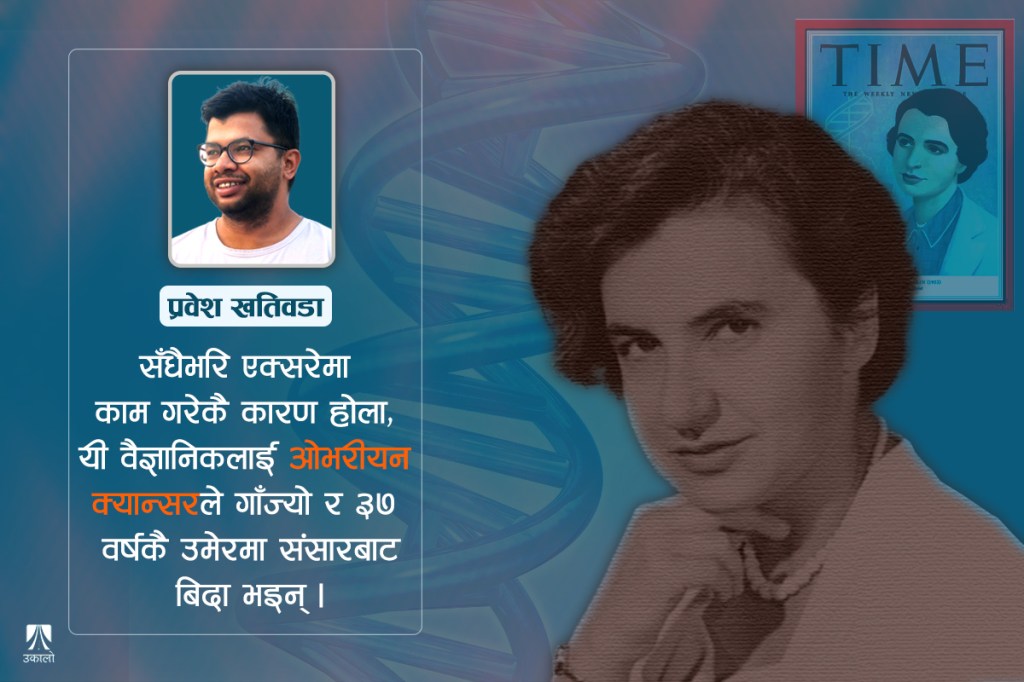
She received little credit for the discovery of DNA until much later, but modern virology holds her in high regard- research on many viruses still builds on her work.
In early 1953, many people believed the American scientist Linus Pauling would be the first to work out the structure of DNA. At the same time in Britain, three scientists – James Watson and Francis Crick in Cambridge, and Maurice Wilkins in London – were also trying to crack it. Their own models had failed again and again, and by January 1953 perhaps even they believed Pauling would get there first.
But in his January 1953 paper on DNA’s structure, Pauling proposed a model very like the one Watson and Crick had already put forward in 1951 and knew did not work. After reading Pauling’s paper, Watson rushed to London to share the happy news with Wilkins.
Arriving in London the next morning, excited, Watson met Wilkins in his lab to discuss what to do next and how to push the research on DNA’s structure forward. It was then that Wilkins showed Watson an X-ray photograph taken eight months earlier by his colleague Rosalind Franklin. It was shown without Franklin’s permission, and without her knowledge.
Rosalind, wronged
The photograph Wilkins showed Watson on the afternoon of 30 January 1953 is what we now know as Photo 51. It may look unremarkable today, but the “X” in Franklin’s Photo 51, and the stripes within it, held a great deal.
The moment he saw it, Watson understood: DNA is made of two strands wound into a helix – a double helix. For Watson and Crick it was a eureka moment. Watson, who hurried back to Cambridge to tell Crick, later wrote that the instant he saw the picture his mouth fell open and his pulse began to race.
Two days later, from 2 February, Watson and Crick restarted their work on DNA’s structure on the basis of Photo 51. By 7 March they had built the model of DNA we know today. On 25 April their paper announcing the structure appeared in Nature under the title “Molecular Structure of Nucleic Acids.”
Franklin, who took Photo 51, had meanwhile left King’s College London for Birkbeck College in 1953, on the condition that she would no longer work on DNA. The head of the King’s laboratory was John Randall, but Wilkins had long treated her as his assistant rather than his colleague. Years later, Watson’s memoir The Double Helix caricatured her as a difficult, combative woman who could not work with others or interpret her own data – a portrait that shaped how the world saw her for decades.
At Birkbeck, Franklin spent the last five years of her life (1953–1958) using X-rays to study the structure of viruses. Working mainly on RNA viruses, she worked out the structure of the tobacco mosaic virus in a short time and began studying the polio virus.
Though she received little credit for the “discovery” of DNA until much later, modern virology holds her in high regard; research on many viruses still builds on her work. Perhaps because she had worked with X-rays all her life, she developed ovarian cancer in 1956, and on 16 April 1958 – sixty-seven years ago today – she died at just 37.
Today Watson and Crick are known everywhere as the scientists who discovered DNA. In 1962, the two of them and Wilkins shared the Nobel Prize for it. The Nobel Prize still cannot be shared by more than three people, and it is still not awarded after death – and women, especially those no longer living, are still passed over.
In 1953 Watson and Crick only built a model of what DNA looks like. Wilkins played a supporting role, providing further X-ray evidence for their model. They did not discover the structure of DNA through their own laboratory experiments. They proposed what DNA’s structure must be, based on other people’s research.
Here I try to trace what research on DNA’s structure came before Watson and Crick, and how we arrived at the double-stranded double helix we know today. It is worth noting that Franklin is not the only forgotten figure in this story.
The search
To trace the search for DNA, we have to go back to 1869, to a Swiss scientist who is hardly remembered today: Friedrich Miescher. While studying proteins in white blood cells, he found a strange molecule. Unlike proteins, it had no sulfur, but it was unusually rich in phosphorus.
Because it was found in the cell’s nucleus, Miescher named it “nuclein.” The nuclein he discovered then is what we now call DNA. Miescher wrote that, like proteins, nuclein might come in many forms.
Science and history forgot Miescher’s research and his “nuclein” for a long time. From the early 1900s, nuclein came to be known as “nucleic acid.”
Only many years later did the Russian-born American scientist Phoebus Levene investigate what nucleic acids were made of. In his “polynucleotide” theory of 1919, he proposed for the first time that nucleic acids are strands made of long chains of different nucleotides. The “tetranucleotide” model Levene later proposed for the sequence of nucleotides turned out to be wrong, but his research was important in the early years of the search for DNA.
It was Levene who first said that each nucleotide is made of a phosphate, a sugar and a nitrogen base. He was also the first to show that there are two kinds of nucleic acid, distinguished by their five-carbon (pentose) sugar. He laid the groundwork for understanding that DNA (deoxyribonucleic acid), found in the cell’s nucleus, and RNA (ribonucleic acid), found both inside and outside the nucleus, differ by a single oxygen atom.
Through the research of Levene and his contemporaries in the 1920s and 1930s, these different nucleotides were gradually identified. It became clear that the nitrogen bases inside nucleotides differ, and that nucleotides fall into two groups based on those bases.
One group, the purines, are made of two carbon-nitrogen rings; they include adenine (A) and guanine (G). The other group has only a single carbon-nitrogen ring. These pyrimidines come in three kinds: cytosine (C), thymine (T) and uracil (U). Three of the bases are found in both DNA and RNA, but thymine and uracil differ: DNA contains thymine, while RNA has uracil in its place.
Unpicking the phosphate, sugar and nitrogen base inside the nucleotide was done much later by Alexander Todd. In his work from the 1940s he showed that the phosphate, sugar and base sit in a fixed arrangement, and how one nucleotide links to the next to form a nucleotide chain (a polynucleotide chain).
This link between nucleotides, known as the phosphodiester bond, can be understood like this: the third carbon of the sugar in one nucleotide joins, through a phosphate, to the fifth carbon of the sugar in the next, and the pattern repeats to form the polynucleotide chain. It is because of the way nucleotides join along the strand that a DNA strand is understood to have a fixed direction (5′ to 3′, or 3′ to 5′). Todd was the first to show that a DNA strand is formed as nucleotide after nucleotide is linked by phosphodiester bonds along a sugar-phosphate backbone.
By the 1940s a great deal was known about what DNA – nucleic acid – was made of, but what DNA actually did was still unknown.
The first to investigate whether DNA stores information, as we now know it does, was Frederick Griffith. Working on bacteria in 1928, he found that the ability to cause disease could be passed from one strain of bacteria to another, and suspected that some “transforming principle” was at work.
Building on Griffith’s work, Avery, MacLeod and McCarty took up the question. In 1944 they showed for the first time that it was DNA, not protein, that carried this hereditary information. Their finding was not widely believed at the time, and strong proof would take another eight years.
In 1952, Martha Chase and Alfred Hershey’s research on viruses confirmed that DNA is the carrier of information. They showed that when bacteriophages – viruses that infect bacteria – attach to bacteria, only the viral DNA enters the bacterial cell, yet new bacteriophages are still produced. With other viruses behaving the same way, no doubt remained that the molecule storing information was DNA, not protein.
By the end of the 1940s it was known that DNA is a long strand made of a chain of four different nucleotides (A, T, G, C), and that it carries information. But much was still unknown, including how DNA’s strands are arranged, and whether it has a single strand or more than one.
Talking about DNA’s strands, we must also know about Erwin Chargaff. Chargaff, who began researching DNA after reading Avery’s 1944 paper, put forward in 1950 what we now call Chargaff’s rules: in DNA, the amount of adenine roughly equals the amount of thymine, and the amount of guanine roughly equals the amount of cytosine – so purines and pyrimidines are present in equal amounts, even though the overall composition varies from species to species.
This result, which held not only for humans, hinted that DNA’s strands might somehow be joined as complements of each other. Chargaff was also among the first to suspect that the nitrogen bases might be linked to one another in some way other than the phosphodiester bonds described above.
Writing later about Avery, Chargaff said that Avery had given us the first text of a new language, and shown us where to look for it – and that he had gone looking. But he, too, was unable to show how A and T, and G and C, pair up (A–T, G–C).
How these nitrogen bases complement each other so that two strands fit together was still unknown. That is the answer Watson and Crick found.
Linus Pauling, mentioned at the start, was another giant alongside Chargaff. Few people did as much as he did to understand how atoms and molecules fit together. He also discovered that proteins form an “alpha helix.” I once read that Pauling, busy at the time investigating how sickle-cell anaemia arises, later regretted not having worked harder on DNA.
He was among the first scientists to suggest that because the strands and bases are complementary, genetic material could pass from one cell to another and from one generation to the next. Speculating about how genes copy themselves, he said in the late 1940s that if a structure consisted of two complementary parts, each part could serve as a template for making the other, and together they would produce a copy of the whole.
Many people believed Pauling would find DNA’s structure first, and Watson and Crick thought so too. But Pauling saw Franklin’s Photo 51 only much later – and his DNA model was born without it.
Watson, Crick, Franklin and Wilkins
Putting it all together brings us to April 1953, when the structure of DNA was announced in Nature. The same issue carried three papers side by side: Watson and Crick’s model, a paper by Wilkins, Stokes and Wilson, and a paper by Franklin and Gosling presenting the X-ray evidence, including Photo 51.
But before connecting them all, we cannot leave out Florence Bell, a forgotten scientist whose work links all four of them. Florence was among the first to study DNA using X-ray photographs. It was her PhD work that led Wilkins to begin researching DNA.
The X-ray photographs in her 1939 PhD thesis showed that DNA must be built from an organised, repeating structure, and that it therefore differs from RNA. It was Florence who first showed that the nucleotide bases are spaced 3.4 ångströms (3.4 Å) apart.
Florence ended her 1939 PhD thesis by writing that working out the chemical make-up of genes was a great and urgent task for humankind; that when we understood it, the limits of our thinking would expand beyond imagination; and that in the end we would learn what we ourselves are.
In the course of improving on her blurry photographs came Franklin and her Photo 51. In 1939, before anyone knew DNA had two strands, Florence had also proposed a single-stranded model of DNA. The Second World War halted her work, and few people read her thesis at the time.
Wilkins was among the few who did. Of the four scientists behind the 1953 papers, Wilkins was also the first to work on DNA. At King’s College London, around 1950, he began taking X-ray photographs of DNA using Florence Bell’s methods.
In the spring of 1951, at a conference in Naples, Wilkins showed his X-ray photographs of DNA. Because of those photographs, Watson – an American who had come to Europe as a postdoc – moved to Cambridge in the autumn of 1951 to work on DNA. In Cambridge he met Crick. Crick was twelve years older than Watson, a graduate student few people liked, and was put in charge of the newcomer.
That is how Watson, Crick and Wilkins came together. From late 1951 they began searching for DNA’s structure. Florence’s photographs were largely ignored at first. In a room in Cambridge, Watson and Crick began building models, fitting DNA’s nucleotide bases together like Lego pieces.
At the end of 1951 they proposed their first model of DNA: three strands, with a phosphate backbone in the middle and the nitrogen bases facing outward, paired with one another.
Phosphate carries a strong negative charge. Packing the phosphates together in one place concentrated negative charge and made the model unstable – a problem that kept tripping them up. The chemical structures of the nitrogen bases were also wrong in the textbooks they used, which added to their troubles. Only when Jerry Donohue corrected them in February 1953 did things fall into place – I will come to that shortly.
In January 1953 Pauling put out a model much like Watson and Crick’s failed 1951 model. Watson and Crick saw his model, published in America, on the evening of 29 January through Pauling’s son, who was studying in Cambridge. The next morning Watson was in London to tell Wilkins that Pauling, too, had proposed a DNA model that didn’t work.
Photo 51
In Wilkins’s lab at King’s College, emptied by the winter cold, they were discussing what to do next. Wilkins suddenly remembered a photograph of DNA that Franklin had taken nearly eight months earlier. Franklin was not in the lab that day, and Wilkins brought out a print of the photograph – Photo 51. After seeing it, Watson hurried back to Cambridge to tell Crick. Fearing that Pauling would spot his mistake and correct it, they went back to work just two days after seeing Photo 51.
Franklin had come to King’s College, where Wilkins worked, in January 1951 on a three-year fellowship as an X-ray crystallographer, originally to work on proteins. Soon after she arrived, the head of the lab assigned her to DNA. Wilkins always considered Franklin to be working under him; Franklin preferred to work independently.
Franklin was not the only scientist studying DNA with X-rays. At a time when no one knew what DNA’s structure looked like, the idea that DNA could take different forms was unknown. Franklin and her student Raymond Gosling were the first to discover that DNA exists in two forms, which they named the “A” and “B” forms.
Before them, everyone who took X-ray photographs of DNA had managed only blurry images. Taking a photograph took a long time, and during it DNA would switch between the A and B forms depending on humidity. By controlling the humidity, Franklin and Gosling succeeded in isolating pure fibres of the A and B forms under fixed conditions, and took separate X-ray photographs of each.
Photo 51 was of the B form of DNA. Franklin and Gosling took it in early May 1952 with an exposure of around 60 hours. At the time there was no eureka moment, and few others saw it. Franklin and Gosling then turned their attention to the A form, which gave more detailed data. They were still working on it in early 1953, and when Watson and Crick restarted their work on DNA, Franklin too had returned to the B form – but the work stalled there.
From Photo 51, it became possible to calculate DNA’s diameter (about 20 ångströms), the length of one turn of the helix (about 34 Å), the distance between two nucleotides (3.4 Å), and therefore that each turn holds ten nucleotides. Together with Franklin’s other data, it also supported the idea that the four nucleotide bases sit inside the sugar-phosphate backbone, pairing with bases on the other strand.
Wilkins showed Photo 51 to Watson without Franklin’s knowledge, and she never learned of it in her lifetime. By then she had already arranged to leave King’s for Birkbeck, and the move came with a condition imposed on her: she was not to work on DNA again.
For Watson and Crick, the Photo 51 that Watson first saw on 30 January 1953 was a kind of master key. Only after seeing it could they show how the nucleotides pair as complements of each other (A–T, G–C), how DNA’s regular, repeating structure is formed, and how one DNA molecule becomes two when a cell divides.
Two days after seeing Photo 51 they went back to their research – there was a race to beat Pauling. Within ten days, by 10 February, they had worked out that DNA is made of two strands running in opposite directions (antiparallel). Until 19 February they were stuck on how like might pair with like – purine with purine, or pyrimidine with pyrimidine. Then they met Jerry Donohue, who pointed out that the textbook structures of the bases were wrong and gave them the correct forms.
After Donohue’s correction, the Lego pieces finally fit! Only then did they arrive at the base-pairing model we know today, realising that a purine pairs with a pyrimidine. On that basis, by 28 February they had solved how the bases pair (A–T, G–C). With the solution came the model we know today: a twisting double helix of two antiparallel strands.
The secret of life
On the night of 28 February 1953, Watson and Crick reportedly went to a pub in Cambridge, bought everyone beer, and announced that they had “found the secret of life.” A few days later, on 7 March, they had finished building the complete model of DNA.
The word spread everywhere that Watson and Crick had discovered DNA. The following month, on 25 April, their paper appeared in Nature under the title “Molecular Structure of Nucleic Acids.” Franklin’s own paper, with Gosling, appeared in the same issue – but as supporting evidence, a few pages behind the famous model.
Had Franklin been alive in 1962, perhaps she, rather than Wilkins, would have received that Nobel Prize. We can only wonder now. But history has many Rosalinds – and doesn’t every year’s Nobel Prize show us something similar?
As biology took another leap forward, it seemed that all the secrets of life might now be revealed. But much remained to be discovered, and it was uncovered bit by bit. Which DNA in our chromosomes becomes RNA, and when, is a whole other strange world – one we came to understand much later, and much of which we still do not know. But science keeps searching, aiming to dig deeper and solve more.
How one DNA molecule becomes two, how it passes from one generation to the next, and how DNA makes RNA and RNA makes protein – the “central dogma” – I will try to discuss in another article.
The Orignal Article was Published on Ukalo at Rosalind’s Birthday, 2025. You can Find it here. https://www.ukaalo.com/news/24848/


Leave a Reply