

“It has not escaped our notice that the specific pairing we have postulated immediately suggests a possible copying mechanism for the genetic material.” – Watson and Crick, Nature, 1953
This is also why marrying within the same family and among close relatives is forbidden. The more the parents’ genes match, the greater the chance that suppressed recessive genes appear in the new child.
Originally published in Nepali in Ukaalo on Friday, 27 June 2025 (13 Asar 2082), as “जिन र डीएनएले कसरी आफ्नो प्रतिलिपि बनाउँछन्?”. Translated into English by the author. Read the original: https://www.ukaalo.com/news/26316/
The research that began after DNA’s structure was found
In the early decades of the twentieth century, many people believed proteins stored genetic information. That made sense: the body contains many kinds of proteins, in many different forms, carrying out countless complex tasks. At the time, no one imagined that a simple molecule like DNA, found in the center of the cell, could hold genetic information. Solid proof that DNA, not protein, stores genetic information came only after Martha Chase and Alfred Hershey’s 1952 research on the T bacteriophage, a virus that infects bacteria.
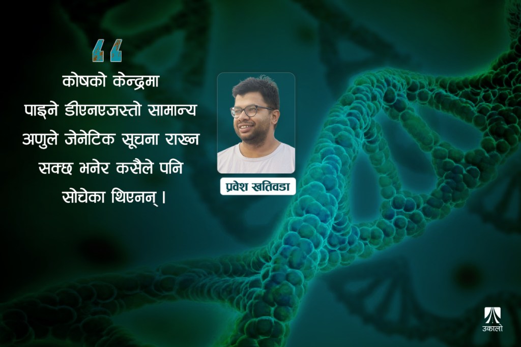
In March 1953, Watson and Crick built a complete model of DNA’s structure. The following month, their paper on DNA’s structure, “Molecular Structure of Nucleic Acids”, had already appeared in the journal Nature. Having shown that DNA has a two-stranded, double-helix structure, they ended the paper with the line quoted at the start. As someone who studied this very subject, I find it more important to understand how DNA copies itself than what DNA’s structure looks like.
Martha and Hershey had given proof that genetic information and the “gene” reside in DNA. Watson and Crick showed what DNA’s structure is. Mendel’s abstract gene had, from then on, found an address in DNA. The search for how DNA copies itself (DNA replication) began after that. Between 1953 and 1957, before proof of the copying process arrived, several different models were proposed.
The first two people to propose a process for copying DNA were Watson and Crick themselves. In May 1953 another paper of theirs appeared in Nature: “Genetical Implications of the Structure of Deoxyribonucleic Acid (DNA)”.
Explaining the structure they had shown two months earlier, they built a model of how, on that basis, DNA could copy itself. The “semi-conservative model of DNA replication” they proposed was later proven true. According to their model, each new DNA molecule contains half (one strand) of the old DNA, while the other half (a new strand) is made as its complement.
(Previous article in the DNA series: Rosalind Franklin: The Forgotten Contributions in the Search for DNA)
For their model to work, the twisted, two-stranded structure of DNA must somehow open up. For DNA to be copied, the job of unwinding those intertwined strands is done by the enzyme helicase. By the time scientists discovered helicase and its role, other interesting models of DNA copying had also emerged. Solid proof of how DNA is copied had to wait until 1957, for Matthew Meselson and Franklin Stahl.
In this article I attempt to discuss the process by which, and the reasons for which, DNA makes an exact new copy of itself. I will also try to connect the research that went into understanding how DNA copies itself. Mistakes can happen while DNA is being copied – and if a mistake happens, what are its effects? I have tried to explain some of this based on my own understanding.
“Gene” and “allele”: Mendel’s research on pea plants
Before understanding how DNA is copied, we need to know the unit that holds hereditary information: the “gene”. The search for the gene takes us back to 1857, to a Hungarian priest, Gregor Mendel, and his research on pea plants.
Mendel, known as the “father of genetics”, chose certain particular traits of pea plants and spent nearly seven years studying how they pass to new generations. His studies showed that in newly born pea plants and in the generations after them, “hereditary traits” pass from one generation to the next bound by fixed rules. It was on the basis of Mendel’s seemingly simple research that we learned each gene requires two “alleles”. Likewise, it was Mendel’s studies that showed that in all sexually reproducing species, one allele must come from the “father” and the other from the “mother” to form a complete gene.
The alleles that make up a gene exist in dominant and recessive forms. The dominant allele keeps the recessive allele suppressed. In other words, if a gene is made of a dominant and a recessive allele, the gene works according to the instructions of the dominant allele.
In his experiments, Mendel assumed that the color of the pea pod is determined by one gene. To create “pure” plants that bore purely green and purely yellow pods, he had them self-pollinate many times. In the “pure” plants bearing green and yellow pods produced by repeated pollination, the color gene is made of two alleles, green-green and yellow-yellow respectively.
Mendel treated the green and yellow colours of the pea pod as the “dominant green” and “recessive yellow” alleles respectively. According to the rule of dominant and recessive genes, a green pod forms whether both alleles are green or one is green and the other yellow; for a yellow pod to form, both alleles must be yellow.
When Mendel cross-pollinated the “pure” plants, the new plants that grew bore only green pods in the first generation. All the new plants of the first generation were born with a gene made of both a green and a yellow allele (green-yellow). After pollinating the plants carrying both green and yellow alleles, however, some pea plants with yellow pods also grew in the second generation. In every four plants of the second generation, the combination of alleles is: one green-green, two green-yellow, and the fourth yellow-yellow.
According to Mendel’s rules, the dominant (green) allele suppresses the recessive (yellow) allele. That is, of every four new plants, three bore green pods. The fourth, with only the recessive yellow-yellow alleles, bore yellow pods.
We still study the rules of heredity that Mendel gave, knowing nothing about DNA or chromosomes, as “Mendel’s laws”. His understanding of dominant and recessive genes is still relevant. Haemophilia, which reached the royal courts of Europe through Britain’s Queen Victoria, spread exactly according to Mendel’s laws. I will bring in the story of haemophilia, known as the “royal disease”, towards the end.
When he presented his research to the Brünn Natural History Society in 1865, no one asked him a question. The minutes of that meeting contain perhaps the most unfortunate note in the history of biology: “Today Father Mendel presented research he carried out over 7 years on pea plants. As there were no comments or questions, we moved on.” The following year, a paper on Mendel’s experiments was published in the same society’s journal: “Experiments on Plant Hybridisation”.
Genes and chromosomes: Mendel’s rebirth and the discovery of the chromosome
Mendel was simply ignored through the nineteenth century. For nearly 35 years no one read “Mendel’s laws” or his research. In 1900, three scientists – Hugo de Vries, Carl Correns and Erich von Tschermak – each carried out experiments like Mendel’s and, while preparing to publish papers with new ideas about heredity, came across that very paper of Mendel’s. It was they who informed everyone that the experiments they had done had already been carried out 35 years earlier by a Hungarian priest. Only then did Mendel begin to be known, as he is today, as the “father of genetics”.
Mendel, who had abandoned science and scientific experiments entirely after the meeting of the Brünn Natural History Society, had already died in 1887. Had Mendel heard of the chromosome, discovered by Walther Flemming in 1879?
As Mendel was being “reborn” at the start of the twentieth century, other scientists had begun to show that Mendel’s laws of heredity could be understood through chromosomes. Flemming had observed that certain fine threads found in the centre of the cell separate, are copied and go into both cells when a cell divides. The name “chromosome” for these units, which under the microscope look like tiny beads on a thread, was given in 1888 by the German scientist Heinrich Wilhelm Waldeyer.
In 1900, Theodor Boveri of Germany, from experiments on sea urchins, first suggested that genes might lie on chromosomes. The next year, the American Walter Sutton showed through experiments on grasshoppers that chromosomes come in pairs, and that Mendel’s genes and alleles can be understood through chromosomes.
The research of these two first revealed that chromosomes are the carriers of hereditary units, the place where genes reside. They were also the first to show that different parts of chromosomes hold different hereditary information.
Chromosomes and genes: the uncertainty between protein and DNA
As early as 1869, Friedrich Miescher had discovered “nuclein”. In 1889, Miescher’s student Richard Altmann gave nuclein, found only in chromosomes, a new “name” – “nucleic acid”. It would still be many decades before nucleic acid became DNA, and chromosomes do not contain only DNA.
Every chromosome contains four kinds of proteins called histones, which surround DNA’s double helix. It is thanks to histones that the long double-helical DNA can be compacted to fit inside a short chromosome. In a sense, histone proteins form an outer shell that protects DNA. Besides histones, chromosomes also contain many other proteins – ones that copy DNA, make RNA from DNA, regulate the histones, and so on.
All the (hereditary) DNA in our body is divided among 46 chromosomes. In everyone, 23 chromosomes come from the father and 23 from the mother. Twenty-two chromosomes are the same in women and men and sit as twenty-two pairs. The twenty-third chromosome, which determines female or male sex, is called the sex chromosome. Its pair is formed a little differently: women have an X-X pair, while men have one X and one short, dangling Y chromosome (X-Y).
Chromosomes contain far more protein than DNA. Proteins are more abundant than DNA, larger, and more complex in structure. It turns out that what Mendel understood as alleles came from these chromosome pairs. It was also understood that for each gene, one copy of the allele/chromosome must come from each of the father and mother, and that chromosomes are distributed in the same way. Yet there was still uncertainty over which molecule of the chromosome – DNA or protein – stored genetic information.
Protein or DNA: the gene finds its address in DNA
Only the research done by Martha and Hershey in 1952 proved that DNA, not protein, holds genetic information.
In 1928, on the basis of experiments on mice, Frederick Griffith first suspected that DNA might carry genetic information. He carried out his experiments on two kinds of bacteria that cause pneumonia in mice: one deadly, the other harmless. When “live harmless” and “dead deadly” bacteria were put together, some “mysterious substance” from the dead deadly bacteria passed into the harmless bacteria and made them deadly too. Griffith named this process “transformation”, but could not reveal what caused it.
Oswald Avery joined DNA research after reading about Griffith’s transformation. To find out what caused Griffith’s transformation, Avery used separate enzymes that break down each of the bacteria’s main molecules (carbohydrate, protein, fat, DNA and RNA). Breaking down every other molecule of the deadly, disease-causing bacteria had no effect. But when DNA was broken down, the ability to cause disease could not pass to the harmless bacteria. Announcing his results in 1944, Avery claimed that it was DNA that held the ability to transform that Griffith had described.
Avery’s results pushed Erwin Chargaff towards DNA research. But even if DNA had the ability to “transform” as Avery and Griffith had thought, few people were ready to accept that genes resided in DNA, or that DNA held hereditary information and passed it to the next generation.
Only the experiments done in 1952 by Martha Chase and Alfred Hershey on the T bacteriophage – a virus made only of DNA and protein, which infects bacteria – proved that DNA, not protein, carries genetic capacity.
For their research, Martha and Hershey labelled sulphur, found only in the virus’s protein, and phosphorus, found in DNA, with different radioactive tags. To find out whether the T bacteriophage’s DNA, its protein or both enter the bacterium to make new viruses during infection, they tracked where the radioactive sulphur and phosphorus ended up in the bacteria.
The DNA at the centre of the virus entered the bacterial cell. The protein in the virus’s outer coat stayed stuck outside the bacterium’s outer membrane. On this basis they concluded that DNA is the hereditary material. Later, once other viruses also showed that only DNA enters the bacteria, there was no longer any doubt that DNA is the unit of hereditary information.
It was only after Martha and Hershey’s experiments that the scientific world began to accept that hereditary units lie not in an abstract “gene” but within the physical structure of the DNA found in chromosomes. It was already known that chromosomes copy themselves during cell division – Martha and Hershey’s discovery gave genetic research a new turn.
From the “invisible gene”, research now began to focus more on how genes and DNA copy themselves. By this time, Chargaff’s rules and Rosalind’s “Photo 51” had already advanced understanding of the DNA inside chromosomes a great deal. At the start of the next year, Watson and Crick were about to reveal both DNA’s structure and the way it is copied.
Watson and Crick: DNA’s two-stranded structure and the semi-conservative model of copying
In April 1953, Watson and Crick showed in their double-helical model that DNA is made of two intertwined strands. On the inside of each DNA strand are the nitrogen bases, and outside them is a sugar-phosphate backbone. These two strands, made of repeating chains of nucleotides, run in opposite, antiparallel directions (one strand 5′–3′ and the other 3′–5′), their nitrogen bases complementing each other and joined by hydrogen bonds. In their April 1953 paper, “Molecular Structure of Nucleic Acids”, they showed that DNA has a structure that repeats according to fixed rules, a double helix like a twisted rope or wire.
The following month, another Watson–Crick paper appeared in Nature itself: “Genetical Implications of the Structure of Deoxyribonucleic Acid (DNA)”. In “Genetical Implications” they explained in detail the DNA model they had given in April, and showed, in a “semi-conservative” model, how DNA and the genes in it could copy themselves on the basis of DNA’s structure. According to Watson and Crick’s semi-conservative model, just as DNA’s two strands fit together, when DNA copies itself the old strands act as templates for the new strands. In Watson and Crick’s model, one strand of the copied DNA is new while the other remains old.
For DNA to be copied, first of all the hydrogen bonds between the nitrogen bases joining the two strands must somehow break, so that the two strands separate. Watson and Crick could not say how DNA’s double helix opens, or what makes DNA’s new strand.
Copying DNA: the different models and proof of the copying process
Closing “Genetical Implications”, Watson and Crick suspected that enzymes made of protein might help open DNA and make and add new nucleotides.
Many accepted their “semi-conservative” model at the time. There were some sceptical scientists who thought it impossible for DNA’s intertwined structure to keep opening and closing. DNA helicase was discovered only much later (1970), and even DNA polymerase had to wait until 1956 to be discovered. Further confusion was added because DNA polymerase can make new nucleotides in only one direction (5′–3′).
One such sceptical scientist was Max Delbrück, who proposed two separate models of DNA copying. The first model, which he proposed in 1953, is called the “conservative” model.
To copy itself, DNA makes a third strand that is not wound into the double helix. On that basis, Delbrück’s model showed that the old cell keeps the old DNA while the new cell receives DNA whose two strands are both new. Having stopped working on this model after hearing a lecture by Watson at the end of 1953, he put forward another model, the “dispersive” model, in 1954.
Insisting that DNA could not possibly separate, in the dispersive model he proposed that new DNA is made by joining small pieces. He tried to show that if every turn of DNA’s double helix (a cycle of 10 nucleotides) were broken in the middle, DNA’s strands would not tangle. Because the DNA strands made this way contain parts of both new and old, it was named accordingly.
Delbrück kept bringing new theories. The enzymes Watson and Crick had imagined had still not been discovered. Watson himself, it seems, had begun to doubt his models. It is said that after arriving at Caltech in the United States in 1954, Watson built three more models there, with the copying nucleotide bases on the outside.
In 1953, while proposing DNA’s double-helix structure, Watson and Crick had written of how DNA copies itself: “As DNA copies itself, each newly formed molecule contains one old and one new strand.”
This hypothesis of Watson and Crick’s was proven by experiment in 1958 by two American scientists, Matthew Meselson and Franklin Stahl. Financial support for their research came from none other than Delbrück. The names of the three models of DNA copying were, in fact, given by Meselson and Stahl.
Meselson and Stahl grew E. coli bacteria for many generations in a medium of heavy nitrogen-16. They later began growing those bacteria – raised in heavy nitrogen-16, with heavy nitrogen-16 in their DNA – in a medium containing light nitrogen-14. The results showed that all the DNA in the first new generation of bacteria was of intermediate density (between light-14 and heavy-16). In the second generation it had split into two layers: one light and one intermediate.
Meselson and Stahl’s result now clearly proved that DNA is copied according to the semi-conservative model. The semi-conservative model of DNA had opened the door to modern genetic studies, but it took several more years to be sure how DNA divides.
Copying DNA: new strands are made by separate processes on the leading and lagging strands
For DNA to be copied, first of all DNA’s double helix must open. At the start of the copying process, the enzyme helicase arrives and attaches. The copying process begins once helicase breaks the hydrogen bonds between the nitrogen bases, separating DNA’s two strands and forming a “replication fork”.
As soon as DNA’s strands separate, two problems arise. If someone tries to pull apart the two strands of a twisted two-strand rope from the middle, the rope resists from the opposite end, doesn’t it? The DNA topoisomerase enzyme relaxes that same resistance from DNA on the far side of the replication fork.
Another problem comes when the separated strands try to rejoin. The strands helicase has separated immediately try to join again by hydrogen bonds. The job of keeping the separated strands in place, not letting them rejoin, is done by SSB (single-strand binding) proteins.
As helicase keeps separating the double helix this way, the replication fork keeps growing. With the strands separated by helicase acting as templates, the process of copying DNA begins. Just as the separated DNA strands run one 5′–3′ and the other 3′–5′, as nucleotides are added along their template, the new DNA strands must be made in the 3′–5′ and 5′–3′ directions respectively.
The enzyme that makes DNA’s new strand, DNA polymerase III, can add nucleotides to a new strand only in the 5′–3′ direction. On that basis, DNA’s old strands are known as the leading (3′–5′) and lagging (5′–3′) strands. On the leading strand DNA can be copied continuously, whereas on the lagging strand it has to be made in stops and starts.
When new DNA begins to be made on both the leading and lagging strands, first of all a small piece of RNA (an RNA primer) must come and attach. The job of bringing small complementary pieces of RNA to the old DNA template opened by DNA helicase, and attaching them, is done by RNA primase. Once the RNA primer is attached, the enzyme DNA polymerase I recognises it and comes to attach.
Two kinds of DNA polymerase are the main ones in DNA copying. The enzyme called “DNA polymerase I”, or “RNase H”, removes the RNA pieces attached at the start. The main role in copying DNA is played by DNA polymerase III.
This is how DNA is copied on the leading strand: a small piece of RNA settles on DNA’s leading strand (3′–5′) in the opposite direction (5′–3′). After DNA nucleotides displace the RNA, DNA polymerase III attaches. On DNA’s leading strand, DNA polymerase III can continuously make new double-stranded DNA in the direction helicase is opening. As soon as the new strand is made, the SSB proteins that attached while it was single-stranded detach and leave.
DNA polymerase III can add nucleotides only in the 5′–3′ direction. On the lagging strand, however, DNA has to be made in the opposite direction to helicase and the leading strand (3′–5′), so that process is not continuous.
To put it briefly: as the helicase enzyme opens the replication fork, new pieces of RNA keep attaching to the lagging strand. DNA polymerase I recognises the attached RNA pieces and comes to remove them. After DNA polymerase I removes the RNA pieces, DNA polymerase III attaches and goes on making small pieces of DNA in the 5′–3′ direction. DNA polymerase can make DNA with new nucleotides, but it cannot join pieces of DNA that have already been made. DNA ligase must then come and join the small pieces of DNA together.
These small pieces of DNA, made on the lagging strand but not yet joined, are called Okazaki fragments. The Japanese scientists Tsuneko and Reiji Okazaki discovered this process of DNA copying on the lagging strand in 1960. Only then was it fully known how DNA copies itself in the semi-conservative model, as Watson and Crick had said.
In bacteria, DNA is short and circular. The DNA copying process, which begins once helicase opens DNA at one spot to form a replication fork, ends with two new DNAs and, along with them, two new bacteria. The place where helicase comes to attach is called the origin of replication, and in bacteria there is only one.
When our DNA, compacted into chromosomes, is copied, however, there can be many such origins of replication, or “replication forks”. As the replication fork opened by the helicase enzyme grows along with the new DNA, it joins a replication fork that began elsewhere. As the replication forks join, the pieces of newly made DNA strand also join. When this process is complete in every chromosome, a new copy of the DNA is made.
DNA mutation: mistakes made while copying, and the mechanisms that correct them
Mistakes certainly happen as DNA polymerase copies DNA by adding new nucleotides. Such mistakes arise from mismatches in base pairing as new nucleotides are added to the new DNA according to the old strand’s template. DNA’s nucleotide bases are bound by fixed rules. If the nitrogen base of a nucleotide in the old DNA is adenine (A), the nucleotide added in the new DNA must have thymine (T); or if it is guanine (G), one with cytosine (C). As DNA polymerase III adds new nucleotides in the 5′–3′ direction, roughly one mistake happens for every ten thousand bases added.
The DNA polymerase III enzyme has its own error-correcting mechanism. DNA polymerase can itself detect and correct, to a large extent, mistakes from mismatched base pairing as it adds nucleotides. While DNA polymerase adds new nucleotides in the 5′–3′ direction, its error-correcting mechanism works in the opposite (3′–5′) direction. If there is a base-pairing mistake, DNA polymerase stops adding new nucleotides, removes the mistake and starts adding new nucleotides again. It is this error-correcting ability of DNA polymerase that reduces the mistakes made while copying DNA to roughly one for every one million nucleotides added.
There is another, additional mechanism that corrects mistakes from mismatched base pairing during DNA copying. There is another group of proteins, called MMR (mismatch repair), that repair mismatches between strands. While correcting mistakes, DNA polymerase may sometimes identify not the newly made nucleotide but the old nucleotide base of the template as the mistake. Such occasional mistakes can occur at one or more than one nucleotide. Such base mismatches are generally seen to occur more often towards the ends of chromosomes.
MMR proteins keep searching for such mismatches and remove the wrong nucleotides. The empty spaces are filled in, corrected, with the help of DNA polymerase. After MMR proteins correct mistakes, now roughly only one mistake occurs for every billion new bases added.
If we joined up all the DNA in our chromosomes, each of our cells contains DNA nearly 3.2 billion nucleotide bases long. By this reckoning, each time a cell divides and DNA copies itself, only about ~3 mistakes might occur.
It is these mistakes during DNA copying that are called mutations. As we grow older, or in unfavourable environments, the mechanisms that detect and correct mistakes during DNA copying grow weaker.
Such mutations in DNA do not arise only during copying. External causes – radiation, carcinogens and so on – can also change DNA’s nucleotides. Such changes in DNA’s nucleotides keep passing into new cells as DNA is copied. If such mistakes occur in germinal cells – sperm and eggs – those mistakes are also passed on to the new generation.
Once it was known how DNA copies itself, it became easier to understand how the genes carried by chromosomes and DNA can pass from one cell to another, or to the next generation. In simple terms, the unit of DNA that can make RNA from DNA, and protein from RNA, is what is called a gene.
DNA contains regions called promoters and enhancers that regulate almost all genes. These can be very far from the gene and much larger. Within a gene too, RNA and then protein are made from a short part of the gene, the “exon”. Most of the gene, the “intron”, only regulates that process. Our DNA is only about 2 percent exon in total, and nearly 24 percent intron. It is because not all the DNA in genes can make protein that our DNA makes only about 30,000 proteins in all. If we joined all the proteins found in the body, they would add up to only about 1.6 million amino acids, or 4.8 million nucleotide bases.
Although all the genes in our body are DNA, it is not true that all DNA is a gene or connected to a gene. Some DNA can make only RNA, from which protein can never be made. There is also “non-coding DNA”, said to make nothing at all. At the tail and tip of every chromosome is a repeating structure of nucleotides called the telomere, which gives stability to DNA and chromosomes. Likewise, in the middle of every chromosome is the centromere, which plays an important role in the division of cells and chromosomes. These themselves, however, cannot make RNA or protein.
Nearly 10 percent of a chromosome’s DNA sits tightly packed and condensed, unusable. RNA can never be made from this DNA, called heterochromatin. Most of the DNA in men’s Y chromosome is heterochromatin like this. The remaining 90 percent of DNA, opened up so that it can make RNA, is called euchromatin.
If a mutation in DNA appears where a gene’s protein is made, its effect will be large. If it falls in a regulatory unit, its effect will be smaller. If it falls in an intron, non-coding DNA or heterochromatin, it will hardly be visible.
The address the gene found in DNA: Mendel’s gene and the laws of inheritance
As Mendel said, the gene was found in the DNA of the chromosome. Now, finally, let us go back once more to Mendel and try to understand genes and alleles in terms of DNA.
Chromosomes sit in pairs: one of the pair comes from the father and the other from the mother. On this basis, every gene has two copies, and that is what, in Mendel’s language, was called an allele. Although both alleles are capable of making RNA and then protein from the gene, generally only one of the two does so. The allele capable of making RNA and protein is called dominant, and the incapable one recessive.
If mistakes made while copying DNA, or from other causes, fall in a gene, they have effects accordingly. Some mutations in DNA can sometimes increase or decrease that ability of the gene. In such a case, the allele with increased ability is given priority, and the proteins that are made come out that way too. In the same way, as DNA is passed down, dominant traits keep appearing while recessive traits may appear sometimes. And sometimes, when a gene’s dominant allele is not strongly dominant, the two alleles may blend to produce an intermediate trait.
To understand dominant and recessive alleles through DNA and chromosomes, it may help to take the example of the “royal disease”, haemophilia, which began with Britain’s Queen Victoria. Haemophilia is caused by mistakes/mutations in the genes needed for blood to clot. This mutation, seen only on the X chromosome and considered a “recessive mistake”, shows no effect when there are two X chromosomes.
Victoria had such a mistake on only one X chromosome. That is, the haemophilia gene on her chromosomes had two alleles. In Mendel’s language, in Victoria the normal dominant allele suppressed the mutation that causes haemophilia. That is why, although Victoria carried the cause of the disease (one allele), the disease did not show in her.
This recessive mistake in Victoria passes to her descendants according to Mendel’s law: her sons have a 50% chance of haemophilia, and her daughters a 50 percent chance of carrying the disease like Victoria. Victoria’s son Leopold died young because of haemophilia. As her daughters married the princes of Europe, the royal disease also reached many of Europe’s royal courts.
Many other genetic diseases can be passed down the same way. Similar changes can occur in the DNA on chromosomes other than X/Y, and can sit in our body as dominant and recessive alleles. If suppressed recessive alleles show up for some reason, diseases like haemophilia may appear.
Some social rules may also have been made because of these same dominant and recessive genes. This is also why marrying within the same family and among close relatives is forbidden. The more the parents’ genes match, the greater the chance that suppressed recessive genes appear in the new child.
In another series, I will try to discuss in more detail the reasoning, causes and effects of marriage and other social rules on the basis of how DNA and genes are passed down.


Leave a Reply